Mitochondria and Brain Disease
By
2010, Vol. 2 No. 03 | pg. 1/1
KEYWORDS:
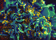
Mitochondria are eukaryotic, membrane-enclosed, 1-10um sized organelles, described as “cellular power plants” as they are responsible for the production of adenosine triphosphate (ATP) and oxidative phosporylation. Signal transduction (buffering and storage of intracellular calcium), control of cell cycle and cell growth, as well as programmed cell death (apoptosis) are other important homeostatic processes governed by mitochondria. It is not surprising therefore that, despite extensive research efforts at elucidating the still un-established pathophysiology of neurological disease, mitochondrial dysfunction is hypothesised to play a substantial role, with their consequent emergence in neuroscience research today. Dysfunction of mitochondrial energy metabolism leads to reduced ATP production, impaired calcium buffering, and generation of reactive oxygen species (ROS) such as superoxide anions, hydroxyl radicals and hydrogen peroxide (Cassarino & Bennett, 1999). ROS are increasingly recognized as playing an important role in neurodegenerative diseases because of their ability to cause oxidative stress and consequently damage cellular contents. Acute exposure to relatively high levels of oxidants, especially in the presence of calcium, can also induce opening of the mitochondrial permeability transition (MPT), an inner mitochondrial membrane residing, voltage- sensitive, non-selective ion channel which opens to pass large molecular weight solutes between the mitochondrial matrix and cytoplasm, enabling the inner membrane (which is normally impermeable) to become permeable, leading to a “large amplitude swelling” (permeability transition) (Kroemer, Galluzzi, & Brenner, 2007). The opening of the MTP is implicated as a mediator of cell death due to a resulting inhibition of the electron transport chain, programmed cell death (apoptosis), oxidative stress, increased leakage of CA2+ currents (which activates apoptotic-inducing factors), as well as cytochrome c release (which activates apoptotic-inducing enzymes and may be a direct consequence of MTP opening) (Norenberg & Rao, 2007). The mitochondrial membrane potential (Ψm), a high -150mV current, is the component of the proton electrochemical potential which determines Ca2+ sequestration and ROS generation. Damage to mitochondrial proteins and mitochondrial DNA (mtDNA) would be expected to decrease mitochondrial bioenergetics and efficiency, as the lack of histones in mtDNA and diminished capacity for DNA repair renders it susceptible to oxidative stress(Lin & Beal, 2006) . Mitochondrial ROS production is intimately linked to mitochondrial membrane potential, such that hyperpolarization increases and promotes ROS production (Valko et al., 2007).It is not surprising that, in adult neurons, which depend primarily on ATP production to meet bioenergetic demands, any compromises in mitochondrial function place neurons at a high risk for both dysfunction and/or death. The association between mitochondrial abnormalities and disease has been known for approximately four decades, with the description of a patient with hypermetabolism and a skeletal muscle biopsy demonstrating large numbers of abnormal mitochondria, a disorder now termed “mitochondrial myopathy” (Cassarino & Bennett, 1999). There exists substantial evidence that mitochondrial dysfunction and oxidative damage may play a key role in the pathogenesis of neurodegenerative disease. Evidence implicating both mitochondrial dysfunction and oxidative damage in the pathogenesis of Alzheimer’s disease (AD), and Huntington’s disease (HD), as well as ischemia and other neurological disorders, continues to accumulate. This review aims to outline the role mitochondria may play in AD, a debilitating CNS disorder for which there is currently no cure, but substantial evidence suggesting a mitochondrial interplay with disease pathogenesis. Mitochondria in Alzheimer’s Disease (AD)AD, the most common form of dementia, is a terminal neurodegenerative disease, neuropathologically characterised by amyloid “senile” plaques (composed of beta-amyloid or AB) and tau-containing neurofibrilliary tangles (NFTs), marked clinically by short term memory loss, confusion, anger and mood swings . Dysfunction of mitochondrial electron transport proteins has been associated with the pathophysiology of AD and Blass and Gibson were among the first who prompted the notion that defective energy metabolism in Alzheimer’s disease (AD) was a fundamental component of the disease (Sullivan & Brown, 2005). The most consistent defect in mitochondrial enzyme activity reported in AD has been the electron transport chain (ETS) carrier cytochrome c oxidase (COX) (Hirai et al., 2001). It has been hypothesized by research groups worldwide that defective mitochondrial metabolism sets up a cascade of pathological events that initiates AD. Disruptions in energy metabolism have been suggested to be a prominent feature, perhaps even a fundamental component, of AD, as depicted from abnormalities in cerebral metabolism, which precede the onset of neurological dysfunction as well as gross neuropathology in AD(Sullivan & Brown, 2005). There is substantial data from positron emission tomography (PET) for example that consistently shows reduced cerebral metabolism in temporoparietal cortices in AD (Sullivan & Brown, 2005). These changes may stem from inhibition of mitochondrial enzymes including pyruvate dehydrogenase, cytochrome c oxidase, and aketoglutarate dehydrogenase. In particular, Amyloid binding alcohol dehydrogenase (ABAD), a mitochondrial matrix-localised enzyme, may be a direct molecular link between amyloid and mitochondrial toxicity. Evidence comes from the finding that amyloid bound to ABAD was found in AD brain mitochondria, and blocking this interaction in vitro suppressed B-amyloid–induced apoptosis and free radical generation in neurons. Furthermore, transgenic mice over expressing ABAD, when crossed with mice over expressing B-amyloid, showed exaggerated oxidative stress and impaired memory. Similarly, α-Ketoglutarate dehydrogenase complex activity is severely decreased in post-mortem AD brain (Hirai et al., 2001). There are strong links between the mitochondrial and amyloid hypotheses. On one hand, mitochondrial dysfunction and oxidative stress may alter APP processing, leading to increased intracellular Aβ accumulation. On the other hand, β-amyloid may cause mitochondrial dysfunction and oxidative stress. A recent paper has shown that that intracellular accumulated β-amyloid precedes both neurofibrilliary tangles and synaptic dysfunction in a transgenic mouse expressing β-amyloid, presenilin, and tau mutations. The effects of crossing mice with a partial deficiency of manganese superoxide dismutase with Tg1995 mice were examined (William et al., 1998). This markedly exacerbated β-amyloid deposition, providing direct evidence of a link between β-amyloid deposition and oxidative damage (Castellani et al., 2002). Studies from our Blass et al have further elucidated the role of mitochondria in AD, by showing that neurons in AD brains accumulate mitochondrial debris in their perikaryon, which results from oxidative damage to mtDNA and mitochondria proteins, and may be related to deficient or defective microtubule metabolism (figure 1). Currently, it is not clear whether oxidative damage to mitochondria leads to a decreased function, or whether a decreased efficiency of the ETS results in excessive electron release and ROS formation. Regardless, the result would be a vicious feed forward cycle where increased oxidative stress would continually reduce mitochondrial bioenergetics. This loss of mitochondrial bioenergetics is coupled with increased oxidative damage and ROS production . A recent study has depicted the relationship between mitochondrial abnormalities in AD and their relationship to oxidative stress, with results showing the same neurons which showed increased oxidative damage in AD had a striking and significant increase in mtDNA and cytochrome oxidase. In addition, morphometric analysis showed that mitochondria were significantly reduced in AD, which together suggests an early association between hallmark neuropathological features of AD and oxidative damage (Hirai et al., 2001). Conclusion and Implications for the FutureMitochondrial defects are now described in a wide spectrum of human conditions, including neurodegenerative and metabolic diseases, aging, and cancer. Further studies examining the importance of mitochondrial pathophysiology in neurodegenerative diseases such as AD and HD may provide important insight into neurodegenerative disease pathogenesis and may indeed provide a target for specific therapies. There is increasing interest in the potential usefulness of coenzymeQ10 (CoQ10) to treat neurodegenerative diseases because CoQ10 administration can increase brain and brain mitochondrial concentrations in brain in mature and older animals. CoQ10 (also known as ubiquinone) serves as an important cofactor of the electron transport chain, where it accepts electrons from complexes I and II, thus serves as an important antioxidant in both mitochondria and lipid membranes. A prior study showed that vitamin E has efficacy in slowing the progression of AD. The antioxidants curcurmin and melatonin exert beneficial effects on amyloid deposition in transgenic mouse models of AD. It is, therefore, possible that CoQ10 might similarly be beneficial in AD. ReferencesCassarino, D. S., & Bennett, J. P. (1999). An evaluation of the role of mitochondria in neurodegenerative diseases: mitochondrial mutations and oxidative pathology, protective nuclear responses, and cell death in neurodegeneration. Brain Research Reviews, 29(1), 1-25. Castellani, R., Hirai, K., Aliev, G., Drew, K. L., Nunomura, A., Takeda, A., et al. (2002). Role of mitochondrial dysfunction in Alzheimer's disease. Journal of Neuroscience Research, 70(3), 357-360. Hirai, K., Aliev, G., Nunomura, A., Fujioka, H., Russell, R. L., Atwood, C. S., et al. (2001). Mitochondrial Abnormalities in Alzheimer's Disease. J. Neurosci., 21(9), 3017-3023. Kroemer, G., Galluzzi, L., & Brenner, C. (2007). Mitochondrial membrane permeabilization in cell death. [Review]. Physiological Reviews, 87(1), 99-163. Lin, M. T., & Beal, M. F. (2006). Mitochondrial dysfunction and oxidative stress in neurodegenerative diseases. Nature, 443(7113), 787-795. Norenberg, M. D., & Rao, K. V. R. (2007). The mitochondrial permeability transition in neurologic disease. [Article]. Neurochemistry International, 50(7-8), 983-997. Sullivan, P. G., & Brown, M. R. (2005). Mitochondrial aging and dysfunction in Alzheimer's disease. Progress in Neuro-Psychopharmacology and Biological Psychiatry, 29(3), 407-410. Valko, M., Leibfritz, D., Moncol, J., Cronin, M. T. D., Mazur, M., & Telser, J. (2007). Free radicals and antioxidants in normal physiological functions and human disease. The International Journal of Biochemistry & Cell Biology, 39(1), 44-84. 
From the Inquiries Journal Blog  ") Monthly Newsletter SignupThe newsletter highlights recent selections from the journal and useful tips from our blog. Suggested Reading from Inquiries Journal
Inquiries Journal provides undergraduate and graduate students around the world a platform for the wide dissemination of academic work over a range of core disciplines. Representing the work of students from hundreds of institutions around the globe, Inquiries Journal's large database of academic articles is completely free. Learn more | Blog | Submit Follow IJ
Latest in Biology |